Learning Objectives
- Vascular malformations include cavernous malformation, telangiectasia, AVM and DVA
- Cavernous malformations have a small risk of spontaneous hemorrhage
- Treatment of these conditions may be observation or surgery
History
KA, a 26 year old female, visits the ER one evening because she has a severe, sudden onset headache. She does not normally have headaches. She describes it as a strong, sharp pain behind her eyes on both sides. She has not had a fever or sinus symptoms. She denies facial numbness or double vision. She feels a little dizzy, but this is much less bothersome than the pain. She denies nausea or light sensitivity.
KA normally takes birth control medication. She has no allergies to medications.
She has a past medical history of one successful pregnancy.
She does not smoke or drink.
A ten system review is negative.
Examination
Her temperature is 99.3 F. Her blood pressure is 125/78 and her pulse is 78. There is no evidence of orthostatic hypotension. She is sitting up on a gurney in the ER. She is awake and alert. Her carotid and cardiovascular exams are normal. Pupillary and funduscopic exams are normal. There is vertical nystagmus when she looks upward. Eye motions are otherwise normal. There is no scalp tenderness. Facial sensation to pin is normal. Movements of facial expression are normal. The cranial nerve exam is otherwise normal. She has normal strength in the arms and the legs on both sides. She has normal deep tendon reflexes. She has normal finger-nose-finger movements. She has a normal Romberg. She has a normal casual gait.
Localization and Neuroanatomy
Pain behind the face is a poor localizing factor. This could indicate the trigeminal nerve ganglia, the cavernous sinuses, the maxillary or ethmoid sinuses, or retro-orbital spaces. Vertical nystagmus is an ominous sign that indicates disruption of the superior gaze centers of the posterior midbrain or the cerebellum.
Diagnosis
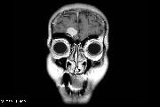
This patient has a severe sudden onset headache. She does not normally have headaches and she does not have symptoms of a benign headache disorder. She deserves a CT scan for evaluation in the ER. The CT scan may miss the midbrain or cerebellar peduncles however, and since these may be affected in this case, a brain MRI would be a better choice.
There are four types of congenital blood vessel malformation that may be found in the brain. These are cavernous malformation, developmental venous anomaly (DVA), arteriovenous malformation (AVM), and capillary telangiectasia. Many of these may cause sudden onset headaches by bleeding. They can be a cause of focal-onset seizures. In most cases, they cause no symptoms. They may be present as a single lesion or as numerous lesions. They are usually incidental findings on a scan done for another purpose.
The cavernous hemangioma (or cavernoma) is a rare cause of headaches. It appears to be an enclosed structure of sinusoidal blood vessels, not well-connected to the rest of the circulation. Cavernomas are usually small and sometimes quite numerous. They pose a risk for spontaneous bleeding- the rate is about 1-2% per year. Cavernomas may be repeat offenders- the risk of bleeding recurrence is 4% or more. Incidental cavernomas are normally candidates for observation, while symptomatic lesions are normally treated with surgery, when possible.
Developmental venous anomaly is a common cerebrovascular lesion. It has been said that 1-2% of people have one of these. It may range in size from a few millimeters to several centimeters. It could be considered a collateral vein that drains a small area. When contrast is given during an imaging study, it enhances similar to other veins. It may not be visible in the absence of contrast. Although they are rarely symptomatic, and they rarely need treatment, DVAs are sometimes treated with surgery.
Another type of vascular malformation is the AVM, or arteriovenous malformation. This is a small bridge from the arterial to the venous circulation, bypassing any tissues in between. If these are large enough, they can interfere with infantile metabolism or development. The shunting of blood from artery to vein results in greater work for the heart, for example. Smaller AVMs may cause headaches, especially if there is a small amount of bleeding associated with them. It is felt the annual risk of bleeding from a cerebral AVM is about 2% per year, for a moderate sized lesion (about 1 inch diameter). Sometimes larger AVMs are surgically removed to prevent this risk. Sometimes an AVM is found to be the cause of a seizure or a progressive neurological deficit.
Capillary telangiectasia is a vascular malformation that is relatively common, but it tends not to be clinically significant. Unlike cavernomas, the capillary telangiectasia has no risk of bleeding. It does not interfere with neurological function or change over time. On MRI studies it appears as a small blush of contrast. This lesion would be surgically removed only in exceptional cases, such as in a case of refractory epilepsy.
Treatment
Congenital cerebrovascular lesions normally require no treatment. Clinical observation is most often recommended for DVA and capillary telangiectasia. Cavernomas and AVMs may be considered for surgical treatment if they are very large or symptomatic.
A variety of techniques may be considered for treatment of cerebrovascular lesions. Traditional open surgery is one option, especially for large lesions. Lesions that cannot be approached by traditional surgery may be candidates for a vascular procedure or stereotactic radiosurgery. The latter has become a popular method for brainstem and cerebellar cavernoma lesions.
Review Questions
- Which of these is a list of cerebral vascular malformations?
a. arteriovenous malformation, aneurysm, stroke
b. developmental venous anomaly, cavernous malformation, capillary telangiectasia
c. capillary telangiectasia, aneurysm, oligodendroglioma
d. aneurysm, developmental venous anomaly, cerebral abscess
e. none of the above - The annual risk of hemorrhage from a cavernous hemangioma is
a. 0-0.5 %
b. 0.5-1%
c. 1-2%
d. 2-4%
e. 4% or more - An asymptomatic cavernous malformation should be treated by
a. clinical observation
b. surgery - An asymptomatic capillary telangiectasia lesion should be treated by
a. clinical observation
b. surgery