Learning Objectives
- Multiple cranial neuropathy is worth considering when cranial nerves that do not co-localize are affected
- There are many causes of multiple cranial neuropathy, including lymphoma, glioma, TB, syphilis, and Cryptococcus
- The treatment of this dire condition depends on the causative agent
History
DT, a 67 year old widow, is brought to your office with symptoms of double vision, facial numbness and facial weakness on the left side. The symptoms have been mildly progressive for the past 3 weeks. She denies pain, trauma or trouble swallowing, but she has opted to stop driving. She has not had headaches. She feels the double vision is improved when she turns her head to the left side. She has been covering her left eye to keep it closed at night time.
She has a past medical history of rheumatoid arthritis and GI bleeding secondary to NSAID medication over usage. She is taking methotrexate and omeprazole. She has no allergies to medications.
There is a family history of seizures in one of her cousins and Bell’s palsy in her aunt.
She does not smoke or drink. She is a retired veterinarian.
Review of systems indicates she had daily joint pain and deformity, but no fevers. She has no trouble with the bowels or the bladder. She has no trouble with breathing or with chest pain. She has no changes in her skin. She has not had bleeding or bruising, fatigue, changes in her weight, or psychiatric symptoms.
Examination
Her pulse is 90, her blood pressure is 118/71, and her temperature is 98.9 F. She is alert, oriented, but she is somewhat older and ill-appearing for her age. Her dress is appropriate. The pupillary and funduscopic exams are normal. Her vision is normal. When she looks to the left, the left eye incompletely abducts. The eye movements are otherwise normal. The left midface (V2) has decreased sensation to pin. The left upper and lower face moves sluggishly, the left eye does not close tightly with effort. The hearing is normal to tuning fork frequencies on both sides. The gag response is weak on the left side, it is normal on the right side. Shoulder shrug and tongue movements are normal. The remainder of the neurological exam is normal.
Localization and Neuroanatomy
This patient has an incomplete, progressive loss of cranial nerve function of V, VI, VII and IX (and possibly X) on the left side. These cranial nerves do not co-localize well, except as pairs. V and VI may localize to the cavernous sinus, VI and VII co-localize to the brainstem roots, VII and IX roughly co-localize either within the pons or along the surface of the ponto-medullary junction. It is difficult to imagine all of them being affected independent of other important structures, such as the corticospinal and spinothalamic tracts. This would be more obvious in situations when cranial nerves are affected on both sides. In this situation one must consider whether multiple cranial nerves are affected after they have exited the CNS. This is a case of multiple cranial neuropathy.
Diagnosis
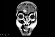
Multiple cranial neuropathy is often a bewildering case to an inexperienced neurologist. The exam is easy enough to observe, the problem is realizing how such a situation could develop. An MRI of the brain with contrast is often helpful for determining or confirming the source of the problem. At times, inexperienced or uninformed radiologists will miss this diagnosis, however, as it can appear vaguely on imaging. It is important to emphasize the exam findings and suspected localizations before ordering the MRI in this case.
There are many cases of multiple cranial neuropathy, some of them are more straightforward than others. Cranial nerves III, IV, V and VI travel through the cavernous sinus on each side, these can be individually affected by pathology in this location (see chapter on cavernous sinus disease). Cranial nerve VII can be affected by Bell’s palsy on both sides. Ramsay Hunt syndrome can affect VII and VIII. Paraganglioma near the jugular vein and carotid artery may affect VII, IX, X and IX.
When cranial nerves are affected in a pattern that does not co-localize, one might consider whether the style of pathology is a coating or disruption of the nerves after they exit the brainstem. Diseases that can cause may be cerebral lymphoma, leptomeningeal gliomatosis, tuberculosis, syphilis, and Cryptococcus fungal infection. These illnesses can otherwise cause significant symptoms and are rarely isolated to the cranial nerves. Penetration of the CNS also portends a negative prognosis. Each of these conditions is treated distinctly. They are often indistinguishable on brain MRI, so CSF studies and sometimes meningeal biopsy are often necessary to complete the diagnostic work up.
In this case, it may be more suspicious that lymphoma is the cause. A small number of patients who use methotrexate develop cerebral lymphoma. Occasionally cerebral lymphoma presents as multiple cranial neuropathy.
Review Questions
- Which of the following cranial nerve deficit symptoms do not co-localize:
a. facial numbness, eye abduction on one side
b. gag response, eye abduction on one side
c. facial weakness, hearing on one side
d. facial weakness, gag response on one side
e. eye lid opening, eye adduction, facial numbness on one side - Causes of multiple cranial neuropathy may include:
a. vitamin B12 deficiency, Ramsay Hunt syndrome, cerebral lymphoma
b. Bell’s palsy, Ramsay Hunt syndrome, Tuberculosis
c. Normal pressure hydrocephalus, Trigeminal neuralgia, Migraine headache
d. Cryptococcus, Toxoplasmosis, Cysticercosis
e. Leptomeningeal gliomatosis, cerebral lymphoma, Epstein-Barr virus
Treatment of multiple cranial neuropathy
a. may be done with intrathecal methotrexate
b. may be done with rituximab
c. may be done with intravenous antiparasitic medication
d. depends on the cause of the neuropathy
e. requires a shot gun approach of multiple systemic medications