Learning Objectives
- Optic neuritis should be evaluated by brain MRI
- Patients with ON respond well to corticosteroid treatment
- Optic neuritis may be a hallmark symptom of MS
- MS symptoms may also be treated with corticosteroids and preventive therapy is indicated
- Most patients with newly-diagnosed MS have a good prognosis
History
A 38 year old woman visits you for symptoms of trouble seeing from her left eye. She felt the symptoms when she woke 2 days ago. There is mild pain when she moves her eye from side to side, but there is no headache. She feels everything she sees is grey and blurred on that side. She feels her pupil is enlarged. Closing the eye seems to correct the visual deficit. There has been no significant change in the vision over the past two days. She visited the local optometrist, who recommended neurology and ophthalmology consultations. She does not have a history of headaches or visual disorders. She denies pain with chewing or opening and closing her jaw.
She has a past medical history of hypothyroidism and gastro esophageal reflux disease. She uses the medications omeprazole and levothyroxine. She has no known allergies to medications.
Family history is negative for autoimmune illnesses. She does not smoke or drink. She is a website designer.
A ten system review of systems is normal, including urinary and bowel function- there is no incontinence, urgency or constipation.
Examination
Blood pressure is 112/66. Pulse is 71. She is a healthy-appearing lady. Heart auscultation is normal. The right pupil reacts to direct light, but not indirect light. The left pupil is enlarged. It reacts to indirect light, but not direct light. The right optic nerve appears normal, the left optic nerve suggests papilledema. She has normal orientation and language. Her fund of knowledge is normal. She has normal visual fields, except visual acuity is impaired in the left eye. Eye movements are normal without nystagmus. Facial sensation to light touch and pinprick is normal. She has normal movement of the muscles of facial expression. Tongue movement is normal. Palate movement is normal. Shoulder shrug is normal. She has normal strength in her arms and legs. She has normal muscle tone. She has normal reflexes in the arms and the legs. The toes are downgoing to plantar stimulation. She has normal sensation to touch, to pin, and to passive movement of the fingers and the toes on both sides. She has normal fine finger movements, normal finger to nose movements, and normal heel to shin movements. She has a normal walk, and she can tandem walk without difficulty.
Localization and Neuroanatomy
There are many possible causes of unilateral visual loss and mild pain. Besides optic neuritis, inflammation or injury to the optic nerve can occur due to vascular disease or inflammation of the optic artery. Retinal detachment or venous occlusion can be a cause. Increased intracranial pressure can affect the vision and result in papilledema. A slow crushing injury of the optic nerve by an aneurysm or glioma can also cause this symptom. Some of these causes can be evaluated by a thorough funduscopic exam, others by MRI of the brain. Many of the alternate causes are rare. This case is suspicious for optic neuritis as the cause, considering the history and exam findings.
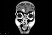
In this particular case, it would be worthwhile to obtain diagnostic testing rapidly and to begin treatment. Laboratory testing to include a complete blood cell count, electrolytes, renal function, and serum sedimentation rate are helpful. They should be normal in a case of optic neuritis. Some providers prefer to evaluate other markers of inflammation. An MRI of the brain is appropriate. This test may demonstrate optic neuritis or many of the alternate causes of this symptom, including those that affect the nerve and the brain.
Treatment
When optic neuritis is the confirmed diagnosis, it behooves the provider to administer treatment. Most neurology providers prefer to treat this condition with intravenous steroids. Some prefer oral corticosteroids. (The medical evidence to help choose between these treatments is not strong.) Fortunately, some cases of optic neuritis improve without treatment, and when they are mild, they may not even be reported by patients. Early treatment of the symptoms is desirable, as they do not respond to treatment at later stages.
Some patients with optic neuritis are found to have a history of other symptoms suggesting inflammation of the central nervous system. Optic neuritis may be a cardinal symptom of a more widespread illness, like multiple sclerosis (MS). Although not all patients with MS will have optic neuritis, and not all patients with optic neuritis will have MS, there is a considerable overlap of the patients affected by these conditions.
Certain MRI findings will suggest an illness like MS, instead of other causes of optic neuritis. These findings are evidence of inflammation in different areas and at different times. This may include regions of contrast enhancement, T2 weighted lesions, or T1 holes. Each of these may represent a demyelinating injury from a different time. Criteria have been developed by neurologists to diagnose MS based on clinical history and MRI findings. At times, MRI of the spinal cord is indicated, as spinal lesions may also occur in MS.
Testing for an illness like MS is not easy or direct. A combination of information helps to build the case. A clinical history, examination findings, and MRI study are a good way to start. When testing by MRI is equivocal, a sample of the CSF can help to make the diagnosis. Certain chemical changes in the CSF occur in the setting of cerebral inflammation. These include the tests for oligoclonal bands, myelin basic protein, and IgG index.
In the case of optic neuritis, patients may feel there is progression of their symptoms over a week, then resolution over four weeks. Treatment with corticosteroids should have a lasting effect; even if the treatment is complete, the healing should continue. The MRI result in an optic neuritis case is helpful for predicting the likelihood of developing MS. When the MRI is entirely normal, there is approximately a 25% chance of developing MS over 10-15 years. If the MRI suggests other symptoms of inflammation, the chance of developing MS is about 75%.
In some cases of optic neuritis, the findings of the MRI study may suggest active MS disease. This might be favored for subclinical areas of inflammation indicated by contrast enhancement. Although these patients will also recover in response to corticosteroid therapy, they should be encouraged to begin treatment to prevent future MS symptoms. This is done with injectable medication, like copaxone, interferon beta 1b and interferon beta 1a. These drugs help prevent other MS attacks and symptoms, some of which may be disabling.
There are also oral treatments to prevent MS symptoms. For the time being, these are limited to patients who have done poorly with injections, or who can afford relatively newer technology that is not yet widely available.
The clinical course of MS varies from patient to patient. Some patients have very little clinical activity, and live a normal life span. Their symptoms may regress for many years, sometimes without treatment. The majority of patients with MS in modern times have a good prognosis. Other patients will have an aggressive clinical course, with multiple attacks, even when treated with preventive medication. They may not recover well from clinical attacks, and they may have a progressive decline in function. Some patients may decline in the absence of attacks, and some may not respond to treatment.
One of the methods used by researchers to monitor clinical progression of MS is by measuring their walking. This may be either a distance covered in a period of time or the time required to walk a certain distance. Some patients will walk more slowly when assistive devices are needed, such as a walker. The progression of MS is also measured by the EDSS scale. This stands for expanded disability status scale. It is not a linear scale. For example, a score of 7.5 indicates no ability to walk more than a few steps, and a score of 5 indicates the ability to walk unaided at about 200 meters. Ability to perform activities of daily living and other symptoms are included in the scale.
When patients are significantly disabled by MS, immune modulatory treatments have less value to their overall condition. Certain alternative medical treatments may be helpful to some patients. Supportive care, nutrition, and maintaining quality of life become more important. A patient with advanced MS with severe symptoms may have the same care requirements as a patient with Alzheimers disease or ALS. Their residual function may depend on the structures of the brain affected by their illness.
Review Questions
- A patient comes to visit you due to loss of vision in one eye. Considering many different possible causes of this symptom, a key part of your diagnostic work up for optic neuritis is:
a. MRI of the brain
b. ocular pressure measurement
c. the clinical finding of papilledema
d. retroorbital pain
e. both A and C - You decide that a patient has recently developed symptoms of optic neuritis. The recommended treatment for this condition is:
a. pain relief with opioid medications
b. laser surgery to reduce pressure in the orbit
c. corticosteroids, either oral or intravenous
d. subcutaneous immune suppressant treatment, such as interferon beta 1b
e. anti-inflammatory medication, such as ibuprofen - A patient with a history of optic neuritis develops symptoms of numbness affecting the left face and arm. They last longer than 24 hours. This indicates the diagnosis of:
a. cerebrovascular disease
b. multiple sclerosis
c. systemic lupus erythematosus
d. complicated migraine headache
e. carpal tunnel syndrome
A patient with newly diagnosed multiple sclerosis has many treatment options. The American Academy of Neurology recommends this as first line treatment:
a. vitamin D 2000 units daily
b. venous angioplasty
c. corticosteroids, either oral or intravenous
d. subcutaneous immune suppressant treatment, such as interferon beta 1b
e. anti-inflammatory medication, such as ibuprofen